Why Advanced Visual Analysis is important
Glaucoma
Several clinic conditions may alter the visual spatial analysis. In order to have a correct diagnosis, it is necessary to take advantage of the most advanced diagnostic technologies.
Let’s discover which are the most recurring pathologies and which diagnostic system we employ to detect them.
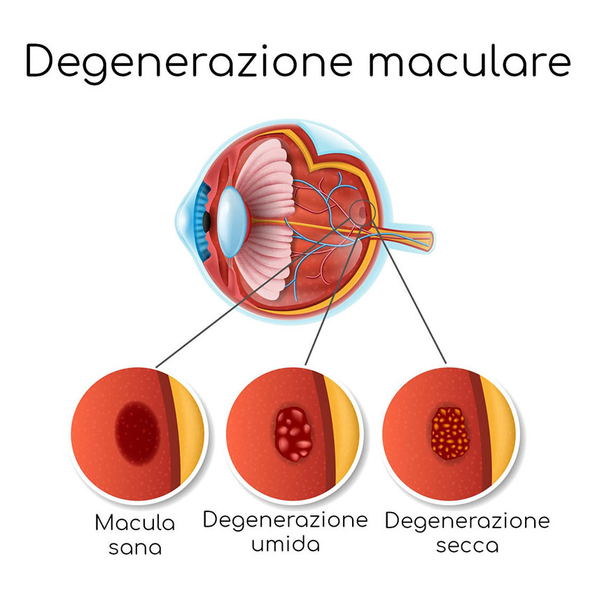
Macular degeneration
It is a retinal disease caused by circulatory factors that affects primarily elders.The reduction of visual acuity makes it impossible to read.
If caught early enough with a regular eye exam, Macular Degeneration can be controlled, stopped or slowed down with a photodynamic laser therapy or intravitreal injections.
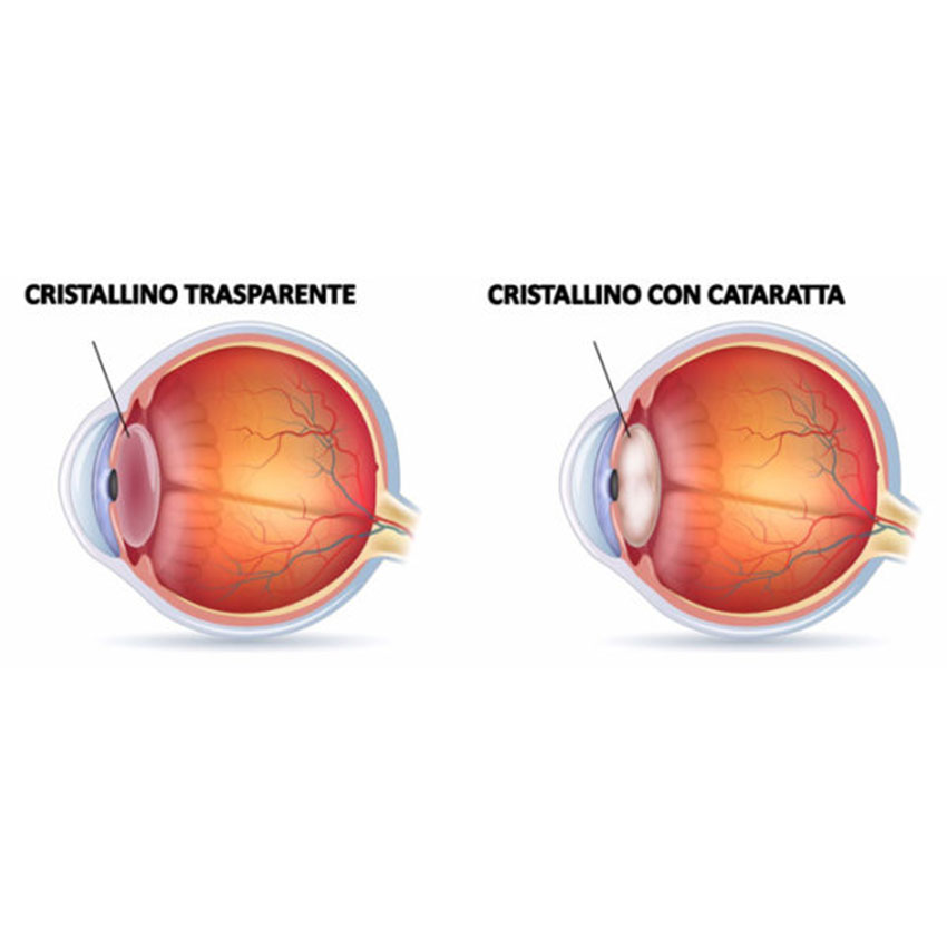
Cataract
Inside the eye there is a lens that focuses clear sharp images on the retina. For people with cataracts these become cloudy. The symptoms of early-stage cataract include mild eye blurriness and cloudiness, with a sensitivity to light and glare, that can lead to a progressive vision loss. After an in-depth evaluation, the ophthalmologist can suggest surgery (Phacoemulsification), usually performed in topical anesthesia and in outpatient basis. It is safe and allows a fast recovery of the visual acuteness in a short time.
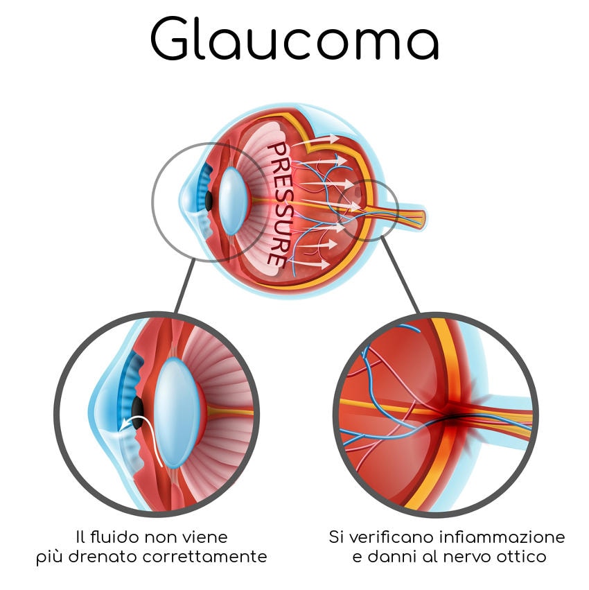
Glaucoma
GLAUCOMA Glaucoma is caused by the increase of acqueous humor intraocular pressure (the transparent fluid that fills the eye). When higher than usual, eye pressure increases significantly the risk of damage to the optic nerve, reducing the visual field. In so doing, it could lead the patient to blindness, without them even realizing.
Generally the therapy consists of instilling eye drops to reduce ocular pressure; laser treatments and surgery are recommended if necessary. However, the simple eye pressure measurement is not enough to diagnose glaucoma, since there are glaucoma with a normal intraocular pressure.
Only the ophthalmologist, with a deep analysis of the optic nerve and with the aid of technologically advanced devices, can detect glaucoma and prescribe the right therapy.
THE IMPORTANCE OF EARLY DIAGNOSIS
Over time, without any treatment, glaucoma can affect vision and eventually lead to blindness. If diagnosed and treated early, the disease progress can be arrested or reduced and the vision preserved. The aim of the therapy is to protect the central vision and the extention of the visual field as much as possible, in order to guarantee the highest quality of life to the patient. This is valid for all the pathologies, from the most aggressive to the less invasive ones. Nevertheless, early diagnosis helps to plan ahead, finding solutions to pathologies before their evolution. As follows, some of our devices to detect them.
Autofluorescence
Thanks to the digital recorded images, Autofluorescence (FAF) is a non-invasive imaging method that allows to identify the retina natural fluorescence derived from the presence of the lipofuscin normally contained in the pigmented layer of retina.
Unlike Fluorescein Angiography or Indocyanine Green Angiography, Autofluorescence exam is performed without any injection.
Why is it so important?
It has been proved that autofluorescence increases in the cells with an accelerated biological activity or in damaged cells, i.e. with a difficulty in working off waste products of the normal metabolic functions. This allows to recognize the retinal damage before it clinically develops.
When is it necessary?
It is useful for the early diagnosis of maculopathies (age-related macular degeneration, central serous chorioretinitis, macular edema, macular hole, Stargardt disease, Best disease) and their follow-up. This allows their precise and non-invasive analysis, especially in patients with allergy to common colorant or with kidney or liver failure, who could not resort to normal angiographic exams.
Optical Coherence Tomography
Optical Coherence Tomography (OCT) is a recent imaging technique, non-invasive, which provides high-resolution images of sections of the human retina "in vivo", allowing the diagnosis, staging and follow-up of numerous retinal diseases. A computer transforms the information obtained into an image on the screen allowing a sort of "in vivo" histological examination of the retina without carrying out any anatomical sampling on the patient.
A false color image is thus displayed in real time which represents the degree of reflectivity of the fabrics placed at different depths: dark colors (blue and black) represent areas of minimum optical reflectivity, while light colors such as red and white define highly reflective areas.
In the early diagnosis of all retinal pathologies involving the macula.
It therefore represents an important step forward in the chapter of preventive medicine.
The OCT offers the possibility to carry out an examination:
- non-invasive
- easily repeatable
- not harmful
- highly precise
When?
This test is suitable for all the pathologies that involve the macula and appear with an immediate alteration of both visual quality (distorted vision, black spot in the centre of the eye) and quantity (from a simple reduction to the complete loss of central vision).
The most frequent pathologies responsible for the loss of central vision can be diabetes, degenerative myopia, age-related macular degeneration or vitreoretinal interface disease (epiretinal membrane, macular hole, vitroretinal cleft). OCT test can identify the more dangerous cases allowing fort he right therapt to follow.

Besides showing visually retina, it is possible to measure the thickness or define the size of a lesion in order to keep it under. In this non-invasive way, you can control the condition of the edema following the therapy and look if the retina is perfectly attached after the detachment surgery. OCT seems to be an irreplaceable test for all the pathologies related to macular region edema, like diabetic retinopathy, forms of vascular thrombosis and choroidal neovascularization. In these particular case OCT scan completes and enforces fluorangiographic examination. Latest devices are endowed with software that analyses the optic papil and the chamber angle. For this reason OCT has become important also for the diagnosis and the follow up of glaucoma.
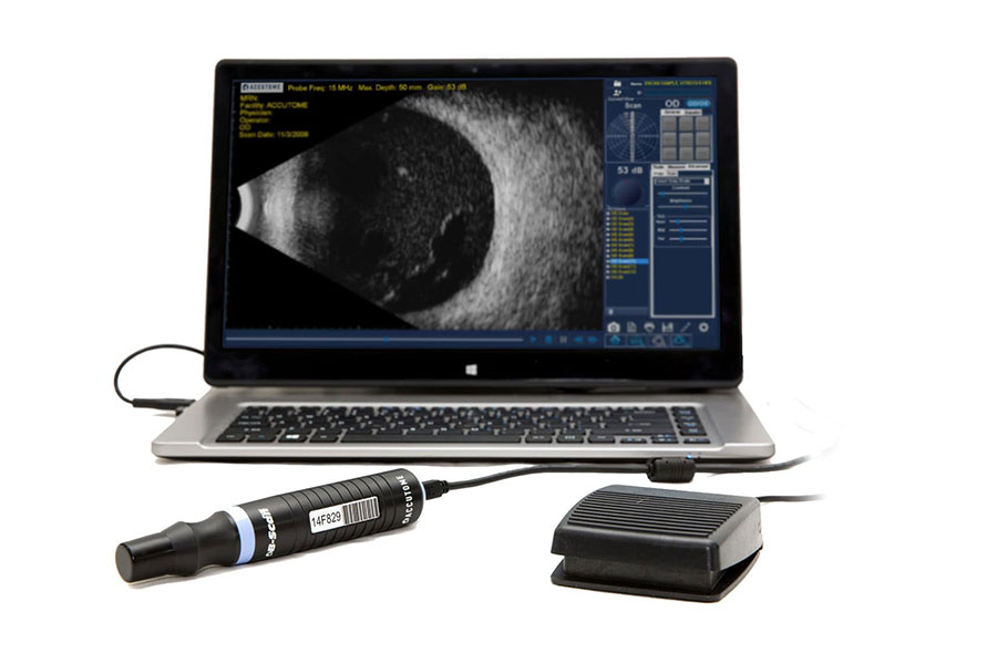
Ultrasound Bio-Microscopy with UBM PLUS®
Ultrasound Bio-Microscopy (UBM) is an high resolution ultrasonic technique that uses ultrasound frequencies up to 48 MHz, providing detailed assessment of anterior segment structures of the eye at very high resolution. UBM allows to measure accurately “Sulcus to Sulcus”, the iridocorneal angle and the anterior chamber of the eyeball.
When is it necessary?
The examination is usually performed on anterior segment structures that normally can not be visualized in another way, as in the case of opaque cornea before the penetrating keratoplasty, when the anterior segment is not known. In case of narrow-angle glaucoma, UBM is very useful for the chamber angle screening, since it allows to verify the correct positioning of the valves and the patency of blebs when the patient is undergoing a filtering surgery. Moreover, it is a method particularly recommended for defining the position of the intraocular lens and for the preoperative measurements in case of phakic intraocular lens implant for myopia. Iris and ciliary body tumors can be visualized and measured in their extention by using the calibers of the tool.
At last, ultrasound bio-microscopy can be very helpful to verify the alterations of the anterior portion of sclera and study accurately ocular traumas (iridodialysis).
Our device
The UBM PLUS® probe, that can be plugged directly into a PC, completes the functions of the B-Scan echograph. The examination is similar to a normal echography, non-invasive and outpatient.
Ecography B-SCAN with B-SCAN PLUS®
B-SCAN ECHOGRAPHY is a non-invasive diagnostic examination that gives detailed bi-dimensional images of the eyeball and the orbit in order to localize and configure lesions.
Our device
Our latest B-SCAN PLUS® portable probe is a practical tool that can be plugged into any laptop or pc.
In addition to the high resolution image (15 micron), with the loss of signal removal as in the common ecographies, and to the possibility to take lasting 34 seconds shot, in order to determine in vivo the histologic aspect of lesions, our device is extremely maneuverable and we can print or send via e-mail the medical report.
Its Smooth Zoom technology allows for 2x full image zoom without distortion or loss of resolution, making this device unique and avantgarde.
When is it necessary?
It is the most important diagnostic test in the presence of opacity in the dioptric media (advanced cataract) to get information about intraocular structures, differently from the other techniques (to detect either retinal detachment or vitreal hemorrhages).
It is useful also in the presence of transparent dioptric media in the differential diagnosis and the evaluation of intraocular tumors dimension.
Regarding orbital level, it allows to evaluate the expansive lesions (tumors), the extraocular muscle (as in the case of thyroid diseases) and the optic nerve in its infraorbital nerve.
Benefits
It’s a painless non-invasive detection that can be performed comfortably in outpatient service.
It is vital for the children study, even the youngest, since it can be performed without being sedated and exposed to radiation. For this reason, echography is ideal for the follow-up of the lesions that need several check-up. Moreover, the medical report is done directly by the ophthalmologist. This is very useful for the radiologist who does not have much experience for evaluating orbital pathologies.
Undoubtedly, along with the common diagnostic radiology exams, it allows the best and the most complete definition and follow-up of the lesions.